When skin cells behave like neurons: The surprising discovery of electrical spikes in epithelia

Author: José R. Pineda got his Ph.D. from University of Barcelona in 2006. Since 2007 he has worked for Institut Curie and The French Alternative Energies and Atomic Energy Commission. Currently he is a researcher of the EHU. He investigates the role of stem cells in physiologic and pathologic conditions.
One of the ideas most of us learn early in biology is that only a few specialized cells can communicate through electrical signals. Neurons do it. Muscle cells do it. Epithelial cells, the cells that form our skin and line most organs, supposedly do not. However, a recent study by Yu and Granick demonstrates that this picture may not be entirely correct. In fact, these seemingly ordinary cells appear capable of generating electrical spikes of their own when tissue integrity is disturbed. Despite epithelial cells have traditionally been viewed as passive structures whose main functions are to form protective barriers, absorb nutrients, and maintain tissue integrity, Yu and Granick challenges this long-standing idea in a striking way. Their work demonstrates that epithelial cells can generate electrical spikes resembling action potentials, revealing a previously unrecognized form of bioelectrical communication that may help coordinate tissue responses after injury .
The finding is surprising because epithelia are among the most abundant tissues in the body. They line the skin, digestive tract, airways, and many internal organs. Although these tissues are known to participate in wound healing and cellular communication, they were not believed to possess the kind of rapid electrical activity usually associated with the nervous system. The new study suggests that this assumption may have been incomplete. Under certain conditions, epithelial cells appear capable of producing coordinated electrical signals that spread across remarkably large distances. To investigate this phenomenon, the researchers employed two different epithelial models: the first model consisted of primary human skin keratinocytes (the major cellular component of the epidermis). The second model used Madin-Darby Canine Kidney (MDCK) cells (a classical epithelial cell line frequently employed in cell biology research). Both cell types were grown until they formed dense, confluent monolayers resembling miniature sheets of epithelial tissue.
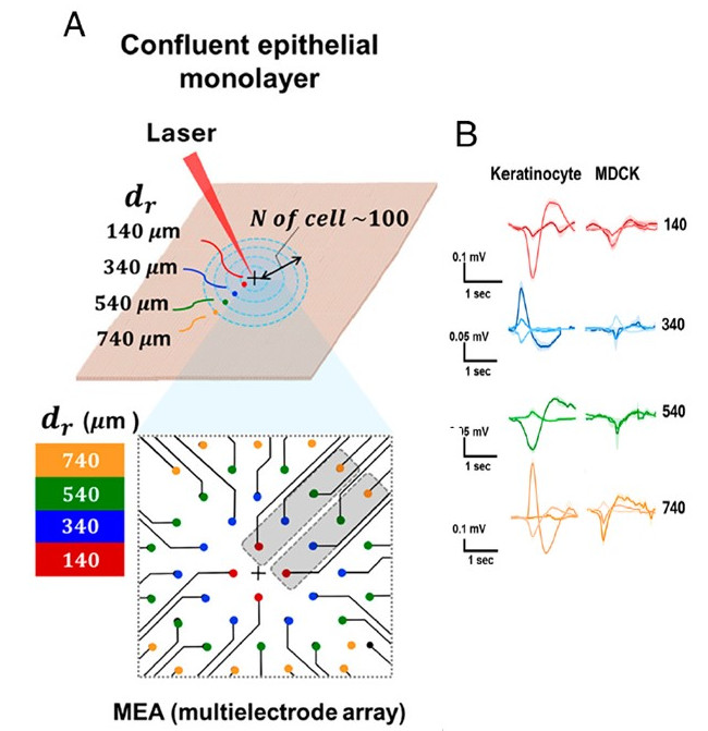
The key experimental tool was a multielectrode array, often abbreviated as “MEA”. This technology can be imagined as a highly sensitive electronic “listening device” containing dozens of microscopic electrodes embedded in a culture surface. When cells generate electrical activity, even extremely small voltage fluctuations can be detected simultaneously at multiple positions. Multielectrode arrays are routinely used to study neurons, but their application to epithelial tissues is relatively uncommon. After establishing epithelial monolayers on these arrays, the researchers created a small and highly localized injury using a focused laser beam. The laser produced controlled damage in a tiny region at the center of the culture while leaving surrounding cells intact. This experimental design allowed the team to observe how cells located at various distances reacted to the injury. Electrical signals were then monitored continuously for one hour. What they observed was unexpected: within the epithelial sheets, electrical spikes appeared shortly after injury and persisted for more than sixty minutes!! These spikes displayed a characteristic sequence of depolarization, repolarization, and hyperpolarization, terms commonly used to describe action potentials in neurons. However, there was one major difference, while neuronal action potentials last a few milliseconds, the epithelial spikes unfolded over one to two seconds, making them roughly one thousand times slower. Despite their slower kinetics, the electrical signals were surprisingly robust, measurements showed that spikes could be detected as far as 740 micrometers from the injury site, a distance corresponding to dozens of cell diameters (Figure 1). Even more remarkably, their amplitude did not progressively decrease with distance as one would expect from a simple wave spreading outward from a central source. In some cases, signals recorded farther away were as large as, or even larger than, those detected closer to the wound. This observation suggested that distant epithelial cells were not merely receiving a weakened signal from injured neighbors. Instead, they may have been generating their own electrical responses as part of a collective tissue-wide process.
Discovering the spikes was only part of the story. The obvious next question was what actually produced them. Were epithelial cells using the same electrical machinery found in neurons, or was something entirely different at work? To answer this, the researchers began dissecting the molecular mechanisms one by one. They began by testing the role of calcium (one of the most important intracellular messengers in biology). Calcium ions regulate a wide range of cellular activities, including contraction, migration, secretion, and gene expression. To determine whether calcium was necessary for epithelial spiking, the researchers added EDTA (a molecule that binds calcium and reduces its availability). Surprisingly, once calcium availability was reduced, the electrical activity virtually vanished!! This immediately suggested that calcium was not just associated with the phenomenon but was an essential part of it. Next, the team examined the contribution of cellular mechanics, because it is known that epithelial cells continuously generate mechanical forces through interactions between actin filaments and myosin motor proteins, a system known as “the actomyosin network”. This machinery allows cells to contract, change shape, and exert forces on neighboring cells. To disrupt this process, the researchers treated cultures with blebbistatin (a selective inhibitor of myosin II activity). Once again, electrical spiking was largely abolished. The similarity between the effects of calcium depletion and myosin inhibition suggested that electrical activity was intimately linked to the mechanical state of the tissue. This observation raised an intriguing possibility: perhaps epithelial electrical signaling is not driven by voltage-gated ion channels (as in neurons), but instead by channels that respond to mechanical forces. To test this hypothesis, the investigators focused on mechanosensitive ion channels, specialized proteins embedded within cell membranes that open when cells are stretched, compressed, or subjected to mechanical stress. Using the inhibitor GsMTx4 (which blocks several mechanosensitive channels including Piezo1), the researchers observed a partial reduction in electrical activity. While spiking was not completely eliminated, both amplitude and coordination decreased substantially. This result implicated mechanosensitive channels as key components of the signaling pathway. To really prove their implication, next the researchers activated them directly using specific pharmacological compounds. One molecule, GSK1016790A, activates the TRPV4 channel. Another, Yoda-1, specifically activates Piezo1. Remarkably, activating these channels was sufficient to generate large electrical spikes even in the absence of any injury. In other words, simply stimulating mechanosensitive channels could reproduce the electrical behavior observed after tissue damage. This provided strong evidence that mechanical sensing lies at the heart of epithelial electrical excitability.
Based on their findings, the authors proposed a positive feedback mechanism: 1) mechanical stress activates mechanosensitive ion channels, allowing calcium to enter the cells. 2) rising calcium levels stimulate actomyosin contraction, generating additional mechanical stress. 3) this new stress then activates more mechanosensitive channels, sustaining the cycle. Such a feedback loop could explain why spiking persists for long periods and why cells distant from the wound remain electrically active. One particularly interesting aspect of the study is its potential relevance to wound healing. Successful tissue repair requires large numbers of cells to coordinate their behavior across considerable distances. Cells must migrate, proliferate, and remodel tissue structures in a synchronized manner. Traditional explanations have emphasized biochemical signaling molecules and calcium waves. The electrical spikes described here introduce an additional layer of communication that may operate on intermediate spatial and temporal scales. According to the authors, these signals could complement chemical and mechanical communication systems, helping tissues organize collective responses after injury. However, the researchers are careful to acknowledge the study’s limitations. Their experiments were performed in cultured epithelial monolayers rather than living organisms. Although the results clearly demonstrate intrinsic electrical excitability in epithelial cells, it remains unknown whether the same phenomenon occurs in intact tissues during natural wound healing. Furthermore, the exact relationship between these electrical spikes and cellular behavior has not yet been determined. It is still unclear whether the spikes actively drive tissue repair or simply accompany mechanical disruption. Additional work involving animal models, voltage-sensitive imaging, and targeted genetic manipulation of mechanosensitive channels will be required to answer these questions.
In conclusion, the study by Yu and Granick reminds us that biology still contains unexpected surprises, even in tissues that have been studied for decades. A simple layer of epithelial cells, once considered electrically silent, may in fact possess its own form of excitability. Rather than transmitting information through millisecond action potentials like neurons, epithelial tissues appear to communicate through slower, mechanically driven electrical spikes capable of coordinating activity across hundreds of micrometers. Whether these signals prove to be a central component of tissue physiology remains to be seen. What is already clear, however, is that the electrical life of cells extends far beyond the nervous system!!
References
- Yu SM, Granick S. (2026) Epithelial cells fire voltage spikes. Proc Natl Acad Sci U S A. doi: 10.1073/pnas.2601750123
The post When skin cells behave like neurons: The surprising discovery of electrical spikes in epithelia appeared first on Mapping Ignorance.

 点赞
点赞 收藏
收藏 点踩
点踩 分享
分享