The Visible Human Project

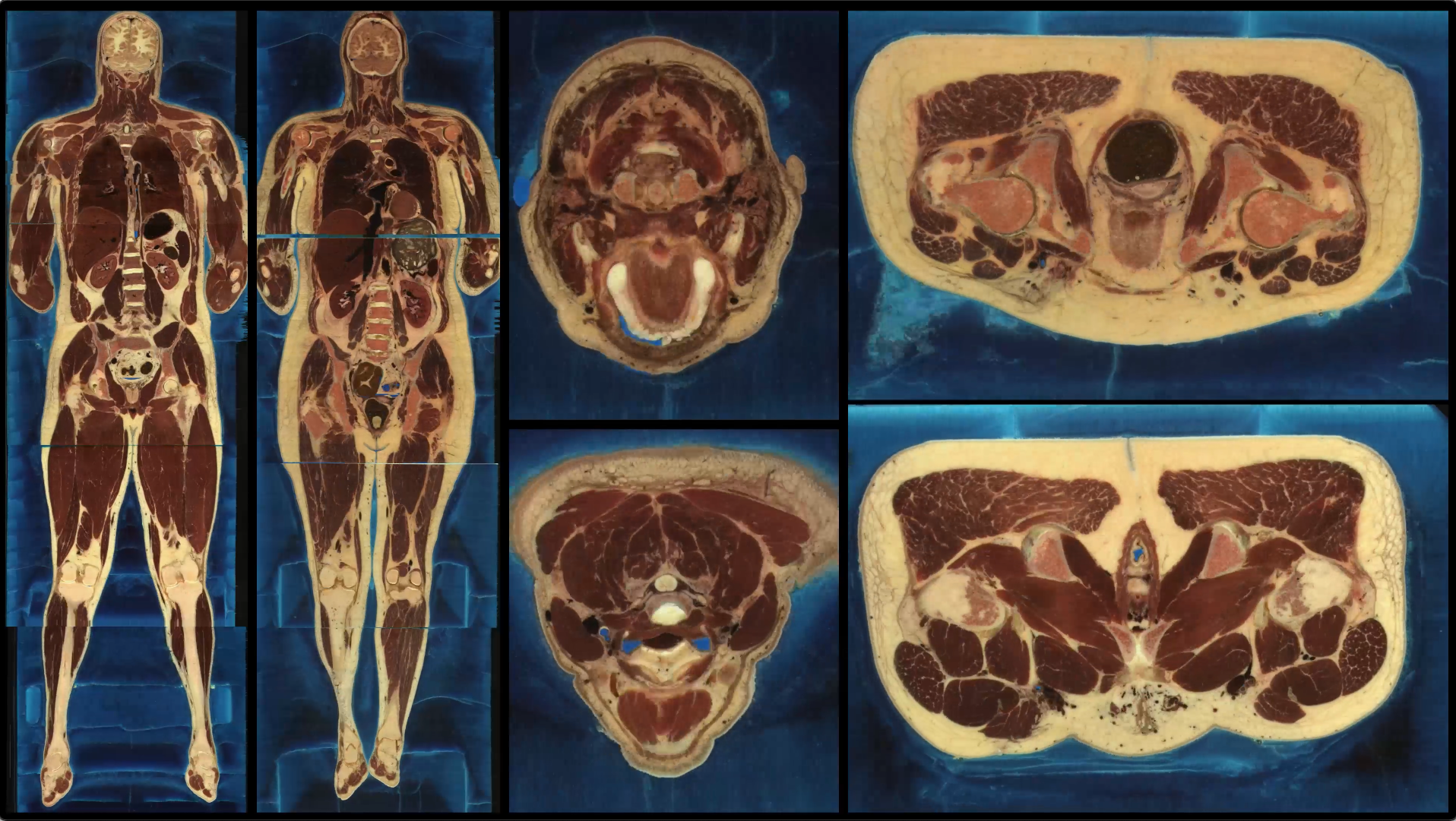
⚠️ Content warning: The images in this post could be upsetting to some readers.
It’s really weird that we don’t know what is going on inside our bodies. Most of us don’t have a clear idea where our various organs are (let alone their actual functions). At the risk of grossing you all out a bit, let’s take a look deep inside the human body. The National Institutes of Health’s National Library of Medicine maintains a valuable and unique dataset that gives viewers an extremely clear picture of what lies beneath our skin.
Created to “produce a system of knowledge structures that will transparently link visual knowledge forms to symbolic knowledge formats such as the names of body parts”, the Visible Human Project is a public dataset that consists of thousands of color photos and scans of two sliced-up human cadavers — one man and one woman.
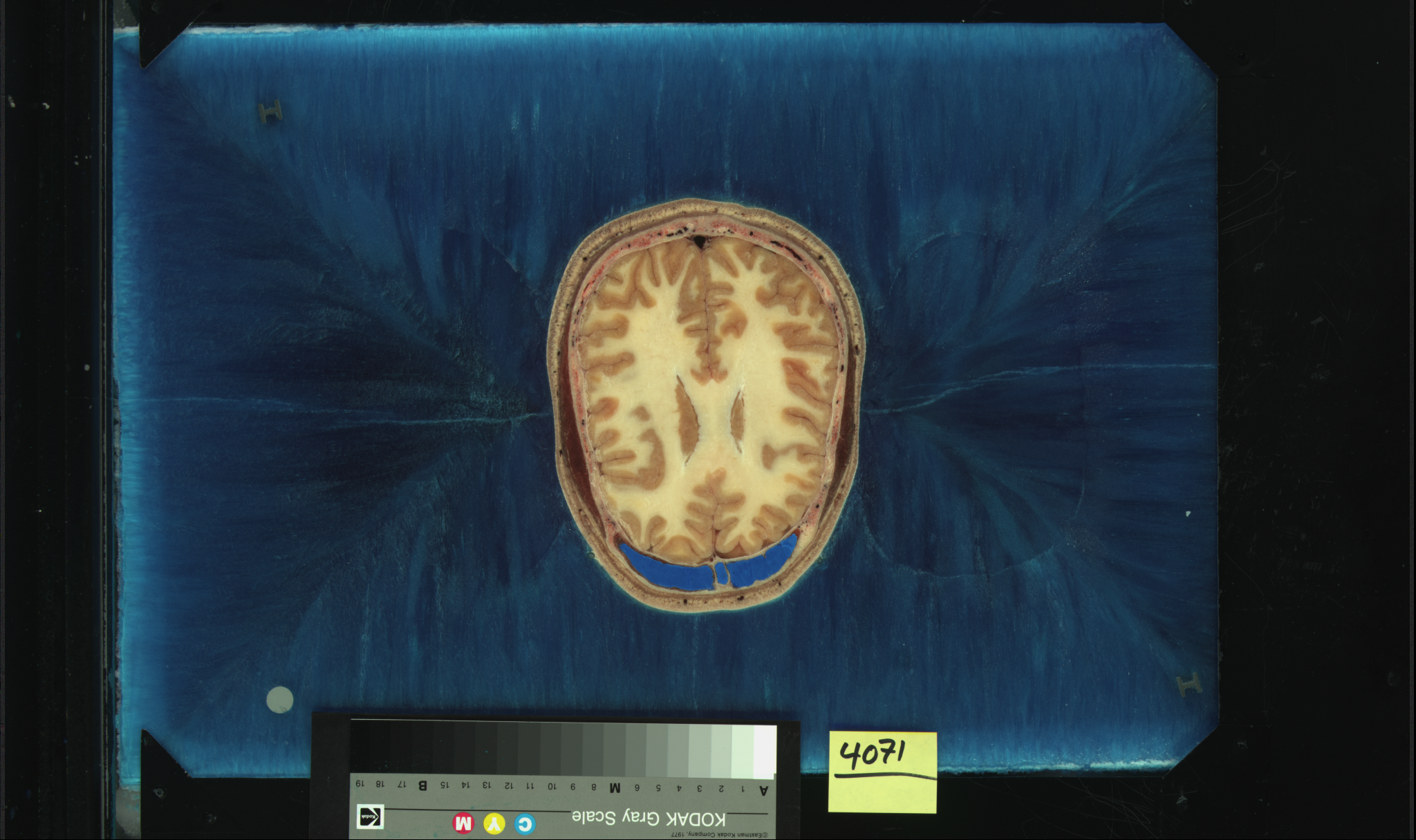
Starting in 1993, researchers at the University of Colorado Anschutz Medical Campus froze the male cadaver, then sliced it up at intervals of 1mm resulting in 1,871 photos. The following year, they did the same with a female cadaver, but at a much higher resolution, slicing at 0.33mm intervals resulting in 5,189 slices. For viewers studying medicine, the frames hold valuable information, allowing granular inspection of how different organs appear in a real body. In addition to the photographs taken by a regular film camera, each body was also captured via MRI and CT scans.
The slices are axial in direction — looking top down on the body. The first images are the very top of the scalp, and the last are the toes and feet. When you watch these frames animated in sequence, the swirls of muscle, fat, and bone explode in abstract patterns. The images are a bit gross at first. Watching them, I feel a mix of fascination and confusion trying to figure out exactly what part of the body I am looking at.
Some parts are very recognizable — the symmetrical hemispheres of the brain are easy to spot, and occasionally you are surprised to recognize the eyes, the heart, or the fingers.
The process was complex: each body was placed in a box, and surrounded with a fast-hardening foam for the scans. After scanning, the box containing the body was frozen. Then the body was cut into four equal-sized blocks. The hardened foam was removed, and each block was placed in a special mold, and filled with a solution of water and gelatin, then frozen to -70C. The frozen blocks were then placed in a custom-made “cryomacrotome” — a specialized device for cutting and imaging specimens slice-by-slice. After each cut, the block was moved to a photographic chamber where the new slice was imaged. A paper describing the process offers more detail:
“In the photographic chamber, each freshly cut surface of the block was flushed with compressed air and examined for defects. Uncut fragments of tendons, fascia, etc., were removed and exposed cavities (e.g. sinuses, trachea, and colon) filled with latex and uncut fragments of tendons, fascia, etc., were trimmed and removed. When the surface was judged to be suitable for photography, it was sprayed with absolute alcohol and surrounded with a black mask, slice number, and a calibrated grey scale.” – “The visible human male: a technical report” *
When the slices are all stacked together, you can also generate a full-body “coronal” view showing slices from the front of the body to the back. This lets medical students examine the cadavers from more than one direction.
Who were these people?
So after staring at every inch of the insides of these two humans, you might ask — who were they?
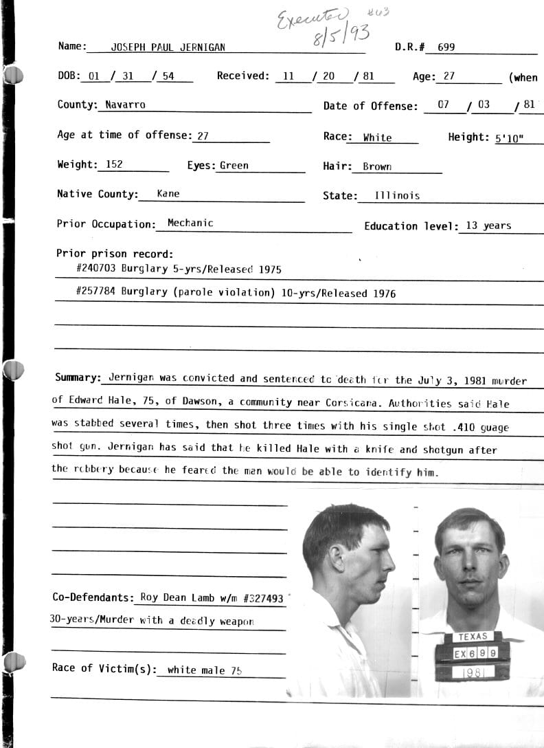
The identities of the donors were meant to be kept private. But back when the project was announced, the identity of the male was quickly discovered: 39-year-old Joseph Paul Jernigan. Jernigan was a convicted murderer who was executed at the Huntsville Unit state penitentiary in Huntsville, TX on August 5, 1993 for killing Edward Hale, 75 of Dawson, TX during a botched robbery in 1981. Jernigan reportedly decided to donate his body to science at the suggestion of the prison chaplain, but it isn’t clear if he knew how it would be used.
Dr. Victor M. Spitzer led the University of Colorado team that created the images. In an 2017 interview with National Geographic, he described the two-year search for the male donor:
“We were looking for a normal body, less than six feet tall, with no trauma or previous surgery. The body had to be fresh. And what could be fresher than someone who is going to die on schedule? For this we needed a source. Which happened to be death row.”
The identity of the woman remains anonymous, but it was reported that she was a 59-year-old white woman who died of a heart attack, who lived in Maryland. Reportedly, the woman’s husband made the decision to donate her body to the project.
Even though the dataset is over 30 years old, it is still widely used. The dataset has been licensed over 4,000 times, and has been incorporated into many teaching aids and training materials.
Willing donor
In 2015, Spitzer’s lab received a new kind of donor. Susan Potter, an 87 year-old Colorado woman who sold flowers near the University of Colorado Health Sciences Center had read about the project, and volunteered her body to be part of the project upon her death. Her years of persistence succeeded, and she forged a friendship with Spitzer. Potter insisted that she inspect the tools and machines that would slice her body up. Potter had survived cancer and other serious illnesses, and her body offered the project a more realistic specimen of a human, complete with the scars and implants from nearly nine decades of a challenging life.
Merch featuring data from our stories
National Geographic followed Potter on her journey in the last 15 years of her life after she was accepted as a donor to the project, with a series of striking photos. Potter’s body bore the marks of a double mastectomy, twenty-six surgeries, and a hip replacement (which had to be removed before the slicing). You can watch a 2018 documentary about Potter and her donation here
Content warning: some of the photos in this video might be disturbing to some readers.
Potter has been immortalized in the project, and advances in technology since the start of the project allowed for each slice of her body to be thinner than a human hair, resulting in over 27,000 images. Extensive interviews with Potter are included as part of her contribution to the project, which will help medical students connect a real person to the images they will be learning from.
In an interview for the project, Potter summed up what she hoped to achieve with her donation:
“It's easy to write a prescription. It's easy to give an injection, or to take you to the surgery and cut you up. That's all easy, there's more to it than that. And that is my project through the visible woman — to bring information and teaching to the medical profession, to the entire healthcare profession.”
*(Spitzer V, Ackerman MJ, Scherzinger AL, Whitlock D. The visible human male: a technical report. J Am Med Inform Assoc. 1996 Mar-Apr;3(2):118-30. doi: 10.1136/jamia.1996.96236280. PMID: 8653448; PMCID: PMC116294)
🙏🏻Did you enjoy this post?
You can subscribe to our newsletter to get future posts delivered to your inbox for free. 👉🏻 📫 .
☕️ Buy me a coffee
Say thanks with a one time tip.
Sharing is caring
📣 If you think your followers or friends may like it, please consider sharing it.
👕🖼️ Buy Beautiful Public Data merch on our shop.
Unique t-shirts and prints inspired by our stories
🙋🏻♀️ If you have any suggestions, comments or requests, please email them to beautifulpublicdata@gmail.com
Thanks for reading!
- Jon Keegan
Bluesky: @jonkeegan.com👨🏻💻Nerdbox
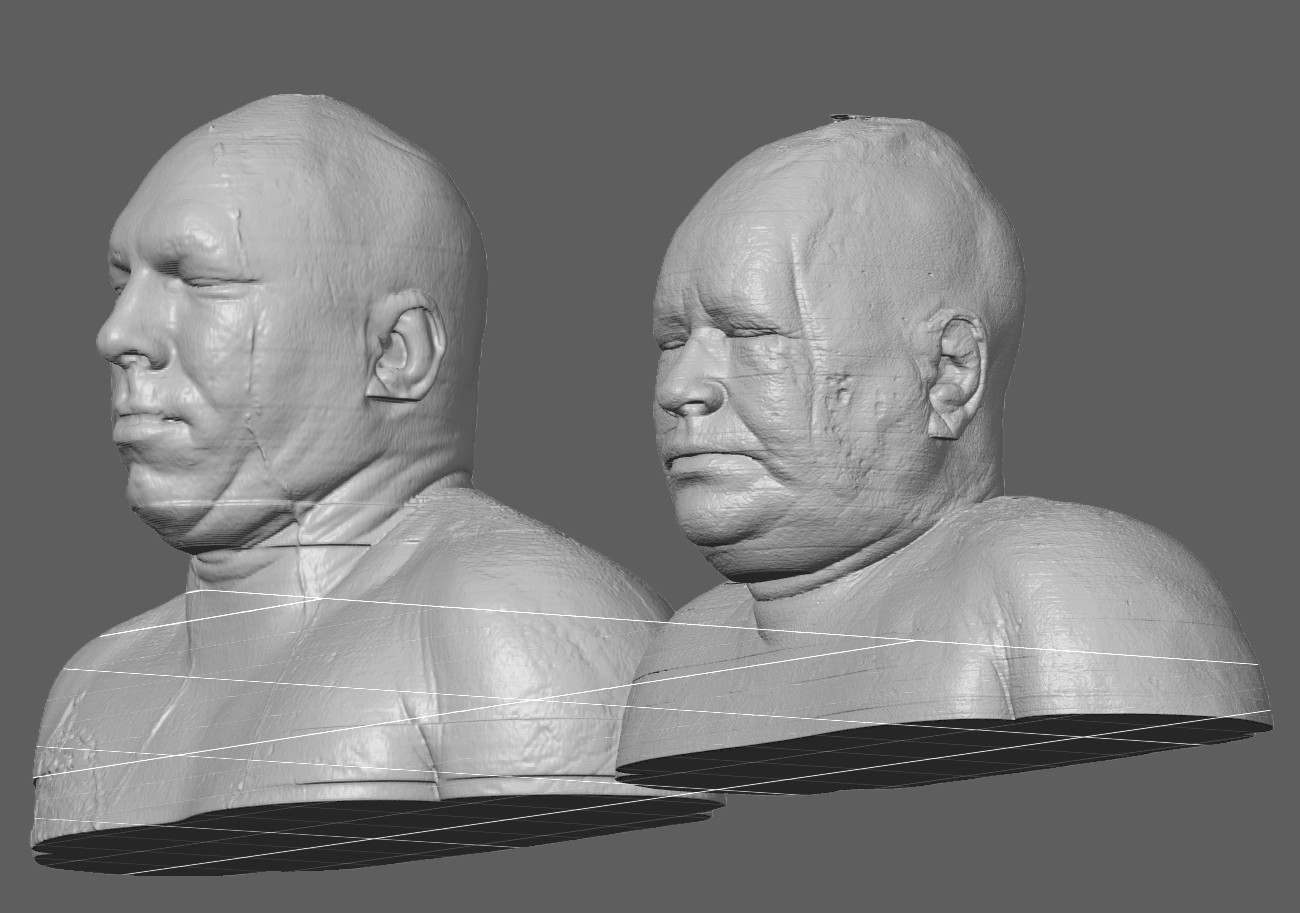
For this post, I used Claude Code extensively to help download and process the imagery from the dataset. I grabbed all of the high-resolution frames from the NLH website, and created animations of them all in sequence using ffmpeg. I then generated a "coronal" animation showing the front-facing full-body view of the slices. I loaded up the STL files in PrusaSlicer and took a screenshot of the models.
To generate the 3-D models of the cadavers, the head and shoulder images were processed to remove the blue support material and generate geometry for 3-D models. Generating the models from the images is a perfect example of a complex image processing task that I would have previously dismissed as not worth the effort, but Claude Code was able to do this on the first try.
You can download the python scripts I used for this post here.

 点赞
点赞 收藏
收藏 点踩
点踩 分享
分享