Monday's Microscope Mystery: What Is This Pink And Red Oddity?

Happy Monday! Sometimes it's nice to start the week by testing your eyes. Because even very ordinary things can look mystifying at a closer angle.
Each week, our team shares a head-scratching microscopic image and asks readers to work out what exactly they're looking at.
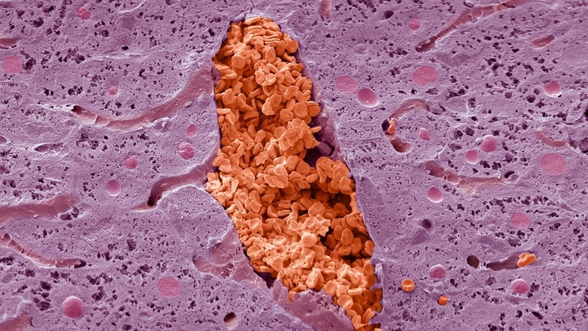
This week's specimen looks almost geological – a jagged reddish-orange seam cutting through pale pink rock, like something you'd find in a cross-section of a mountain rather than the human body.
What do you think you're looking at?
- Click here if you want some options to narrow it down…
- Click here for the answer (spoiler alert, obviously)
There will be spoilers below, you have been warned!
For a visual buffer, here's the most important science news you need to know from last week.
Narrow it down…
Some ideas to narrow down what the image could be of:
a. A kidney stone forming
b. A vein in the liver, packed with red blood cells
c. A crack in tooth enamel
d. A fat deposit in bone marrow
We'll show you the image one more time and will reveal the answer below.
Okay ready?
You sure?
Alright… here we go.
The answer
The answer is B: a vein in the liver, packed with red blood cells.
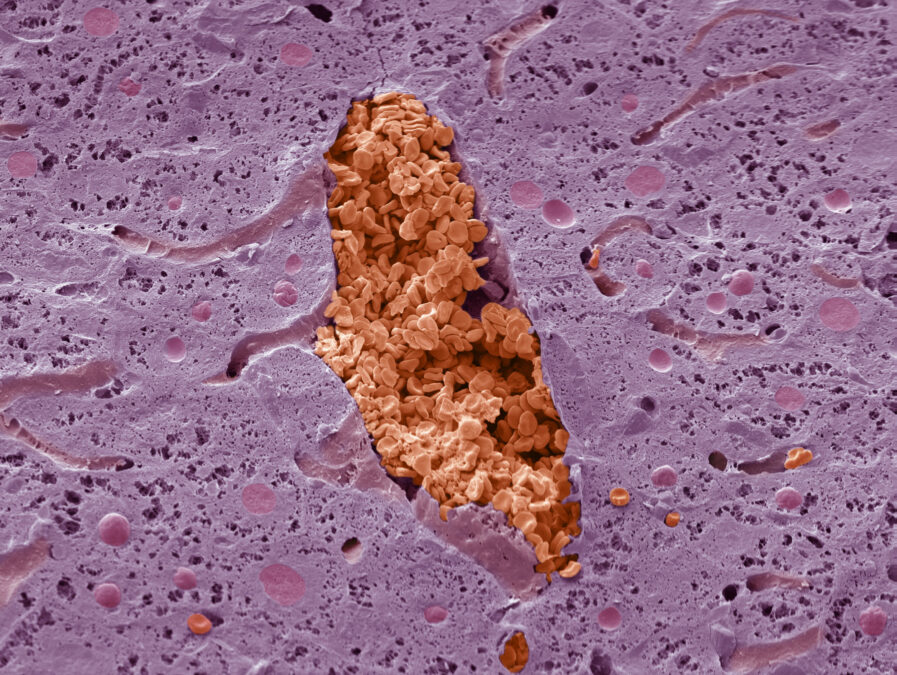
That jagged reddish-orange channel is a vein, sliced through and filled edge to edge with red blood cells, or erythrocytes.
Every red blood cell passing through that vein has already squeezed through liver capillaries barely wide enough to fit a single cell at a time, brushing past hepatocytes on its way through one of the body's busiest filtration systems.

Veins like this one are the return leg of the liver's blood supply, carrying blood that's already dropped off its nutrients and oxygen back toward the heart.
The liver has a dual blood supply: the hepatic artery delivers oxygen-rich blood, while the hepatic portal vein brings blood with nutrients absorbed from the gut. These two supplies mix in the liver's sinusoids before draining out through veins like this one.
The purple tissue packed around the vein is made up of hepatocytes – the liver's main working cells, responsible for filtering toxins, producing bile, and storing energy.
They make up roughly 80 percent of the liver's total volume, arranged in sheets typically just one cell thick so that blood can pass close by every single one of them.
The image was captured using a scanning electron microscope, at a magnification of roughly 200x when printed 10 centimeters (~4 inches) wide, then colorized afterward to make the different structures easier to tell apart.
A scanning electron microscope captures highly detailed images using electrons rather than visible light, so the original is displayed in grayscale.
In real life, blood's red color appears only when oxygen-carrying hemoglobin absorbs and reflects light in the visible spectrum.

 点赞
点赞 收藏
收藏 点踩
点踩 分享
分享